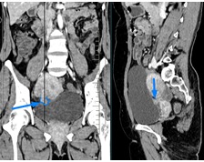
A small focal dome shaped enhancing right posterolateral bladder wall thickening, measuring about 2 cm at the base and 0.9 cm in maximum thickness an apparent small pedicular communication with right anterolateral adjoining myometrial wall of uterus, almost at the site of prior LSCS scar– likely suggestive of a small bladder endometriosis possibly related to prior LSCS scar implantation.
A hypodense heterogeneously enhancing intramural area measuring about 3.3 x 2.6 cm at the right lateral wall of uterus with small contour bulge – suggestive of a fibroid versus focal adenomyosis.
Recommend MRI pelvis, cystoscopy, biopsy