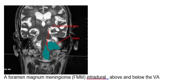
A large extra-axial isointense homogeneously enhancing intradural mass lesion (5.0×3.8×1.9 cm) broad based along the left lateral and anterior wall of foramen magnum, demonstrating intracranial and extracranial components, extending inferiorly up to C2 vertebral level, approximately 1.8 cm below the level of foramen magnum (the component superior to the level of foramen magnum measures 1.7 cm in supero-inferior length) – consistent with foramen magnum meningioma (intradural)
The mass is displacing the medulla to the right, causing marked extrinsic medullary compression. The inferiormost component of the mass is extending posterior to the cervical cord and displacing the cord anteriorly. The superior intracranial component of the mass is indenting the left cerebellar tonsil.
The inferior component of the mass is predominantly posterior to the dentate ligament.
The central portion of the mass at the level of foramen magnum is completely encasing the V4 segment of left vertebral artery causing significant luminal narrowing (the mass is extending superior as well as inferior to V4)
No evidence of perilesional vasogenic edema. No evidence of bony erosions.
Age related volume loss with marked periventricular, subcortical white matter chronic small vessel ischemia with lacunar infarcts in bilateral capsuloganglionic regions, corona radiata , thalami
Recommend HPE correlation, follow up