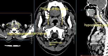
Extensive asymmetrical circumferential heterogeneously enhancing mass lesion predominantly epicentered at the posterior wall of hypopharynx involving bilateral pyriform fossa, aryepiglottic folds, extending caudally to involve the postcricoid region and proximal cervical esophagus measuring 7.5 cm in superior inferior extent with maximum thickness of 1.7 cm – represents carcinoma of the hypopharynx – post cricoid region with proximal esophageal involvement
Segmental narrowing of the hypopharyngeal, proximal esophageal lumen.
Early transmural infiltration of the mass lesion into bilateral tracheoesophageal groove and the prevertebral space at left paramedian aspect. No evidence of vertebral erosion.
The mass is indenting posterior margins of bilateral thyroid lobes with slightly obscured fat planes.
Extensive necrotic metastatic lymphadenopathy in bilateral level I, level II, level III and level IV cervical compartments, largest lymph node measuring 4.2 x 2.6 x 2.5 cm in right level IV
Enlarged necrotic lymph nodes in right level VI compartment and bilateral lateral retropharyngeal spaces in the upper cervical region (*Nodes of Rouvière).
Emphysematous changes in the lungs bilaterally